Electrochleography measures the electrical responses of sensory cells in the inner ear in response to a sound stimulus. In particular, it measures the electrical activity of the hair cells of the cochlea, which are responsible for translating sound signals into electrical signals, and that of the auditory nerve.
When a sound is presented to the ear, it causes movements in the inner hair cells of the cochlea, which generate an electrical response. This response can be recorded by electrodes placed in the external auditory canal or in contact with the eardrum. Electrocleography measures the amplitude and latency of this electrical response.
Electrocleography is indicated in the following situations :
Assessment of deafness : electrocleography can be used to determine the cause of deafness in a patient, helping to determine whether the deafness is caused by damage to the cochlea or damage to the auditory nerve.
Diagnosis of Ménière’s disease: electrocholleography can help diagnose Ménière’s disease, an inner ear disorder that causes vertigo, tinnitus and hearing loss.
Assessment of tinnitus: electroacleography can help determine the cause of tinnitus.
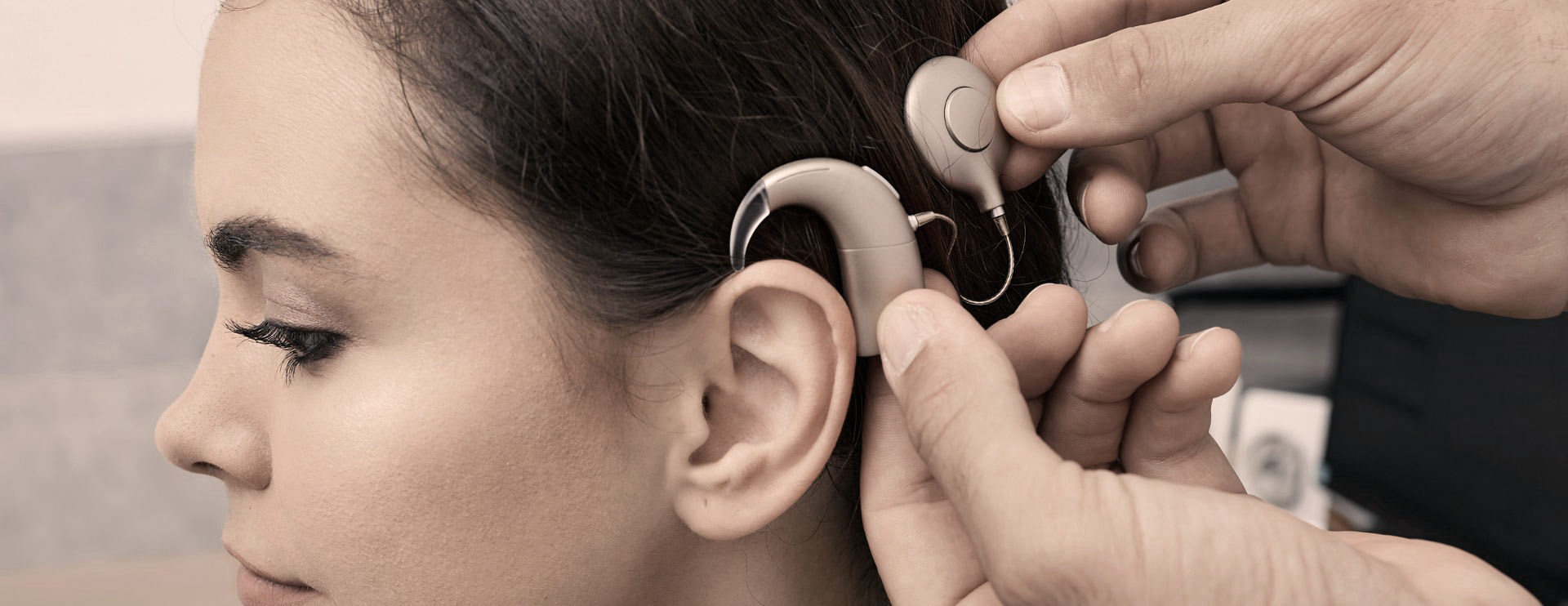
Finally, electrocochleography can be used to assess the feasibility of a cochlear implant and to fine-tune the device.
This examination is usually complementary to other tests of the auditory system, particularly auditory evoked potentials.
Electrocochleography is an objective test that records the electrical response of the inner ear (cochlea) and the auditory nerve when sounds are presented. It helps us to understand how sound is converted into a nerve signal.
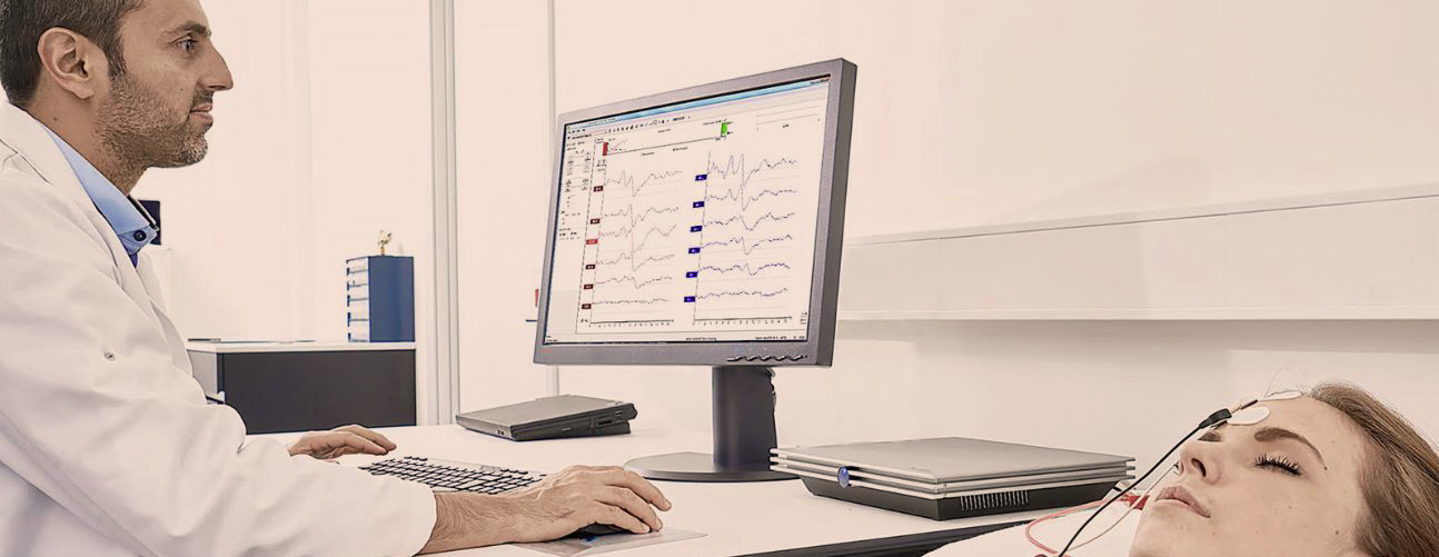
For electrocochleography, small electrodes are placed on the skin (forehead/behind the ear) and a thin electrode is placed in the ear canal (sometimes in contact with the eardrum, depending on the indication). Sounds are delivered to the ear through an earpiece or headphones, and the device records the strength and timing of the response.
The ear-canal method is well tolerated: you will simply feel the earpiece in place.
The eardrum-contact method is carried out under local anaesthetic and may cause minimal, temporary discomfort.
If you have an ear infection, discharge from the ear or a perforated eardrum, the test will be adapted or postponed.

Electrocochleography usually takes 30 to 45 minutes, depending on the protocol (ear canal or eardrum) and the ear being tested.
Electrocochleography may be carried out as part of a hearing loss assessment, to pinpoint more precisely where the hearing impairment lies (cochlea or auditory nerve). It may also be performed when Ménière’s disease/endolymphatic hydrops is suspected, or as part of a cochlear implant plan.
Cochlear implant plan: it helps to assess feasibility and to optimise the settings.
In children, ECochG is mainly reserved for the assessment prior to cochlear implantation. It is carried out by the team that will perform the implantation.